Imaging tests have become routine in A&E, in health checks and even in quick consultations.
But a new study pours cold water on this apparent technological comfort.
The number of CT scans has surged, bringing faster and more accurate diagnoses. Researchers now point to a possible hidden cost: a significant increase in cancer risk over the coming decades, linked to the radiation from these scans.
A study that raises an amber warning
Published in JAMA Internal Medicine, the study analysed the use of CT scans in the United States in 2023. There were 93 million scans carried out in around 62 million people.
Using risk models already employed in radiation protection, the authors project something concerning: this volume could be associated, in the future, with around 103,000 additional lifetime cancer cases among these patients.
The researchers estimate that, if today’s pace continues, CT-related cancers could account for around 5% of all new cancer diagnoses each year.
This does not mean every CT scan causes a tumour, nor that everyone scanned will develop problems. The logic is probabilistic: the higher the cumulative exposure to ionising radiation, the greater the chance of DNA changes in cells-changes that, in some cases, can develop into cancer.
CT is not the villain, but it has a hidden cost
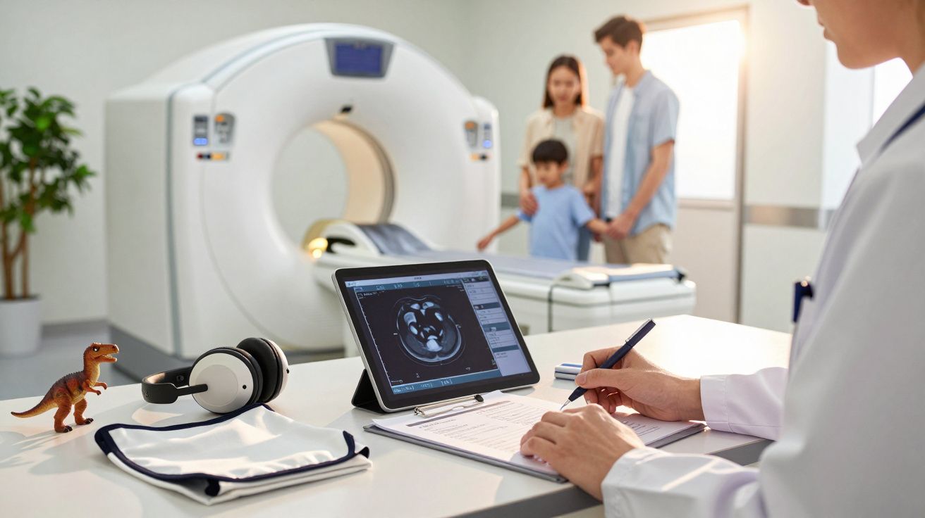
A CT (computed tomography) scan uses X-rays from multiple angles to create cross-sectional “slices” of the body. It shows internal organs, bones, blood vessels and soft tissues clearly, and is often decisive in urgent situations.
It helps detect:
- tumours in various organs;
- clots and emboli;
- internal bleeding;
- deep infections;
- complex fractures.
In cases of stroke, severe trauma or suspected pulmonary embolism, for example, the scan can literally change the course of care within minutes. In these situations, the benefit tends to outweigh any theoretical future cancer risk by a wide margin.
The debate is not “CT: yes or no?”, but “when, for whom, and at what dose?”
Why children are more concerning
Another key point in the study is the difference in impact by age group. Children and teenagers appear as the most vulnerable groups to the cumulative effects of radiation.
Two reasons explain this:
- a young body is still developing, with a higher rate of cell division;
- there is more life ahead, increasing the window in which DNA damage can evolve into cancer.
In adults, the most critical scans, according to the analysis, are those of the abdomen, pelvis and chest. In children, the main focus of attention is head CT, which is common in minor head injury assessments and neurological investigations.
How different organs factor into risk estimates
| Region scanned | Most commonly cited cancers | Who is of greatest concern |
|---|---|---|
| Chest | Lung, breast | Adults and teenagers |
| Abdomen and pelvis | Colon, bladder, blood cancers | Adults |
| Neck | Thyroid | Children and young people |
| Head | Cumulative dose to brain tissue | Children |
Radiologists challenge the broad alarm
Medical societies responded quickly. The American College of Radiology, representing radiologists in the United States, said there is no direct, definitive proof that CT scans cause cancer in humans at the doses used in routine clinical practice.
Specialists note that much of the evidence comes from extrapolations of data from atomic bomb survivors and radiation-exposed workers-contexts very different from modern medicine.
Radiologists argue that, when appropriately indicated, CT avoids unnecessary surgery, speeds up diagnosis and improves survival, reducing preventable deaths.
The disagreement centres on statistical interpretation. Public health researchers warn about population-level impact, while clinicians and radiologists emphasise case-by-case decision-making in the clinic or A&E.
Limit or optimise? The challenge of “less, but better”
Between fear of radiation and the risk of missing a diagnosis, an intermediate approach emerges: not simply doing fewer CT scans, but making them more appropriate and safer.
Measures that are already gaining momentum
- Review protocols: reduce radiation dose without losing image quality, with adjustments by weight, age and body region.
- Avoid repeat scans: check whether the patient has had a recent CT before ordering a similar one.
- Prioritise other methods: where possible, use MRI or ultrasound instead of CT, as they do not use ionising radiation.
- Justify each request: record clearly why the scan will influence clinical management.
Hospitals that adopt optimisation programmes can significantly reduce the average dose per scan. In some cases, radiation savings exceed 30% without losing information that is clinically relevant.
What patients can ask before having the scan
People on the receiving end often wonder: “Should I refuse a CT scan?” In most urgent scenarios, that doesn’t make sense. The bigger issue is elective scans-health checks or repeated scans within a short interval.
Asking questions doesn’t get in the way of care-and it can help prevent automatic requests made out of habit rather than need.
Useful questions to ask your clinician include:
- “Will this scan change what you do in my treatment?”
- “Is there an alternative with less radiation, such as ultrasound or MRI?”
- “Do you have access to images from scans I’ve had recently?”
- “Can the dose be adjusted for my age and weight?”
Why the topic matters in the UK too
Although the study uses US data, the direction of travel is familiar in the UK: growing access to scanners, the marketing of private health-check packages, and pressure for rapid diagnosis.
Across both NHS and private services, patients’ imaging histories are not always easily shared between organisations. That can lead to unnecessary repeats-because earlier reports aren’t available, images can’t be accessed, or teams simply aren’t communicating.
Commissioners and NHS managers are also part of the equation. Fewer unnecessary scans mean better use of resources and, at the same time, lower population exposure to cumulative risk.
Terms worth a quick explanation
Computed tomography (CT): an imaging test that uses high-energy X-rays and computers to produce detailed cross-sectional images of the body.
Ionising radiation: radiation that can remove electrons from atoms and molecules. This can damage DNA and, in some cases, contribute to tumour formation.
Cumulative dose: the total radiation received across multiple tests over a lifetime. A single scan usually carries a very low risk, but repeated scans over time can increase the probability of harm.
Practical scenarios: when the risk is worth it-and when it deserves debate
Imagine someone with severe chest pain, shortness of breath and suspected pulmonary embolism. A contrast CT pulmonary angiogram is close to the standard approach. Without it, a clinician could miss the diagnosis, fail to prescribe anticoagulation, or even proceed to unnecessary procedures. The future cancer risk associated with that scan is likely small compared with the immediate danger.
Now consider a healthy young adult having an annual check-up with a “package” of tests including a whole-body CT scan, without any specific symptoms. In that context, the balance shifts: the concrete benefit is debatable, and the unnecessary radiation exposure repeats year after year.
Between these extremes are grey areas: non-specific chronic pain, dizziness, minor head knocks in children. This is exactly where stricter protocols-and good communication between clinician, radiologist and patient-can help prevent overuse.


Comments
No comments yet. Be the first to comment!
Leave a Comment